Responsible for the facility
Main research areas
Measurements of the texture in samples of structural materials, as well as in geological and biological samples
Facility layout
Main view of facility 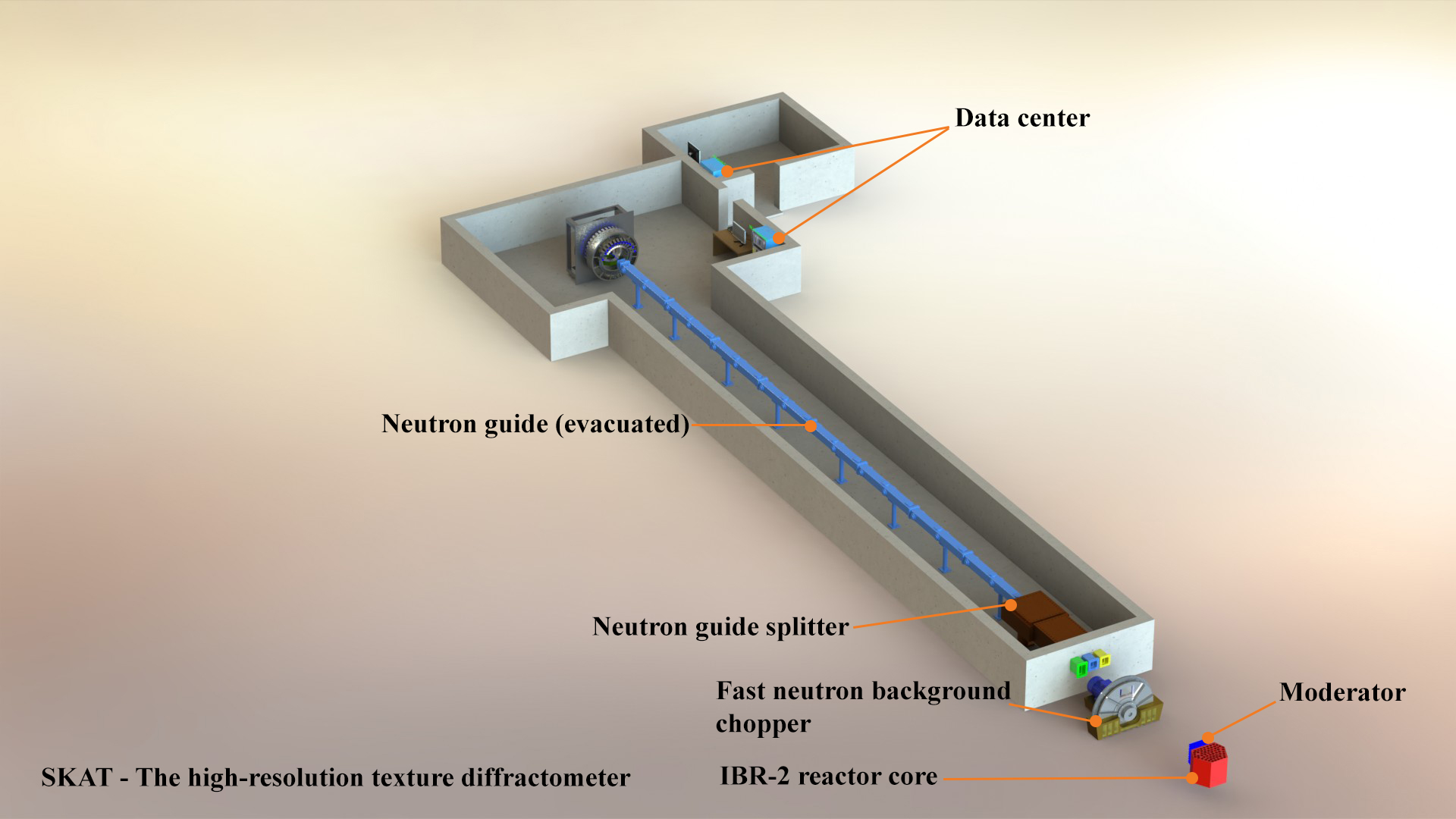
Sample environment 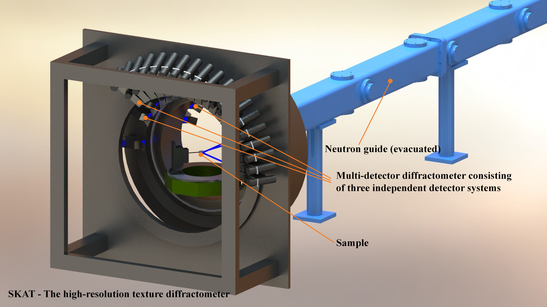
Description of SKAT
The SKAT facility was designed for the measurement of the crystallographic texture that is an ensemble of polycrystal grain orientations. Interest in the research of crystallographic textures relates, on the one hand, to the fact that the texture occurs during the production and increase of polycrystals and on the other hand, to the fact that the texture determines the anisotropy of the physical properties of materials. Traditionally, textures are measured using X-ray diffraction. However, for coarse-grained rock samples, structural materials and biological objects with grain sizes on the order of several millimeters, the use of neutron diffraction is the only way to provide the grain statistics required for quantitative texture analysis. This is due to the greater penetrating power of thermal neutrons compared to X-rays.
The SKAT neutron diffractometer consists of a neutron beam chopper, an evacuated neutron guide, a beam collimation system and a ring detector system consisting of 19 detectors. Detector-collimator complexes are installed on a Debye-Scherrer cone around the incident beam at a scattering angle 2θ = 90°. In addition, it is possible to measure at other scattering angles 2θ = 65° and 2θ = 135°. Measurements at the same scattering angles lead to the same position of the same diffraction peaks for all detectors, so there is no need to introduce λ- and θ-corrections. The detector system, positioned at a scattering angle of 2θ = 90°, allows to carry out texture measurements with high resolution in the range of interplanar distances up to dmax = 5.0 Å. The goniometer angle of 45° is optimal for installation of the equipment for in situ experiments.
Main characteristics
| Path length | ~ 104 m | ||
| Scattering angles 2θ | 65° | 90° | 135° |
| λmax | 7.0 Å | ||
| 2θ-related parameters | 2θ = 65° | 2θ = 90° | 2θ = 135° |
| dmax | 6.5 Å | 5.0 Å | 3.9 Å |
| Best resolution Δd/d | 6.2·10-3 | 5.0·10-3 | 3.1·10-3 |
| Path length after scattering on the sample | 1.10 m | 1.00 m | 0.95 m |
| Neutron guide |
Cross section: 50 mm (w) × 90 mm (h) Radius: 13400 m Coating material: natural Ni (m = 1) |
||
| Detectors |
Set of 19 He3 detectors P = 4.5 bar Ø = 60 mm |
||
| Collimators |
Gd-coated soller collimators Angular dispersion: 18' / 45' Cross section: 55 × 55 mm2 |
||
| Sample positioning | 3-axis geometer | ||
| Data processing | SONIX software PC running Windows OS | ||
Sample environment
- A device for heating the sample up to 1000 °C will be designed.
References
- Ullemeyer, K., Spalthoff, P., Heinitz, J., Isakov, N.N., Nikitin, A.N., Weber, K. The SKAT texture diffractometer at the pulsed reactor IBR-2 at Dubna: experimental layout and first measurements, Nuclear Instruments and Methods in Physics Research, 1998, A412, 80-88.
- Keppler R., Ullemeyer K., Behrmann J. H. & Stipp M, Potential of full pattern fit methods for the texture analysis of geological materials: implications from texture measurements at the recently upgraded neutron time-of-flight diffractometer SKAT, Journal of Applied Crystallography, 2014, 47, 1520-1534
- Lychagina, D. Nikolayev Quantitative comparison of the measured crystallographic textures, Journal of Applied Crystallography, 2016, 49(4), 1290-1299.
- Nikolayev, T. Lychagina, A.A. Zisman, E. Yashina Directly verifiable neutron diffraction technique to determine retained austenite in steel Advanced Engineering Materials, 2017, DOI 10.1002/adem.20170559
- Lychagina, D. Nikolayev, A. Sanin, J. Tatarko, K. Ullemeyer Investigation of wheel steel crystallographic texture changes due to modification and thermo-mechanical treatment, IOP Conference Series: Materials Science and Engineering, V. 82, Proc. XVII Int. Conf. on Textures of Materials, 2015, http://dx.doi.org/10.1088/1757-899X/82/1/012107
- Nikolayev D, Lychagina T, Pakhnevich A. 2019 Experimental neutron pole figures of minerals composing the bivalve mollusc shells // Springer Nature Applied Sciences V. 1, https://doi.org/10.1007/s42452-019-0355-1.