M. Arzumanyan, K. Z. Mamatkulov
D. S. Zakrytnaya, E. Arynbek, E. M. Demina, A. A. Shutikov
Over the past few years, Raman scattering spectroscopy has become a powerful diagnostic instrument in Life Sciences, in particular, in research on programmed cell death.
Neutrophils are the most abundant white cells in the human blood and consist an essential part of innate immunity, providing a rapid response to microbial invasion. NETosis is a process of programmed death of neutrophil cells, distinct from apoptosis or necrosis. In addition to its role as a first line of defense in the innate immune system, dysregulation of NETosis relates to the pathology of various diseases such as rheumatoid arthritis, systemic lupus erythematosus, psoriasis, thrombosis, atherosclerosis and cancer. The mechanisms of activation and the underlying cascade processes of NETosis depend on the specific stimulus. Neutrophil extracellular traps (NETs) are produced using neutrophil granulocytes and consist of decondensed chromatin decorated with antimicrobial peptides. They protect the organism from foreign bodies and are released in the presence of various activators, including pathogens, inflammatory mediators, chemical triggers, ultraviolet radiation (UV) and others.
Kinetic analysis using highly sensitive vibrational spectroscopy, aimed at carrying out this research at FLNP, allowed to reveal the evolution (increase) of the citrulline peak in the low-frequency range of the Raman scattering spectrum of neutrophil cells within 30-40 minutes (Fig. 1) after the onset of the inflammatory process that can be classified as an early diagnosis of NETosis [1].
Fig. 1. Low-frequency region of the Raman spectrum of neutrophils: the intensity evolution (increase) of the citrulline line indicating pre-activation of NETosis 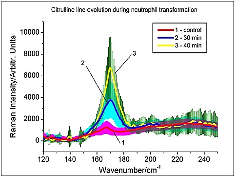
Since the peak with a Raman shift of ~170 cm-1 almost lacks in inactivated neutrophils and increases significantly after activation, it can be assumed that it is due to the accumulation of citrulline in the cell, the spectrum of which also contains a characteristic peak at about 170 cm-1. As a rule, citrulline almost lacks in human cells, since it is not among the 20 basic amino acids from which the proteins of our body are made. However, it is known that during the process of NETosis, citrulline can be produced primarily by transforming histones that are the most essential structural chromosomal proteins within the nuclei of neutrophils.
A little-studied aspect of NETosis is its activation by light, in particular, by UV and visible radiation that is currently the subject of wide research at FLNP, using Raman spectroscopy combined with fluorescence microscopy (Fig. 2).
Fig. 2. Fluorescence microscopy: imaging of neutrophil extracellular traps (NETs) formed under UV(A) radiation 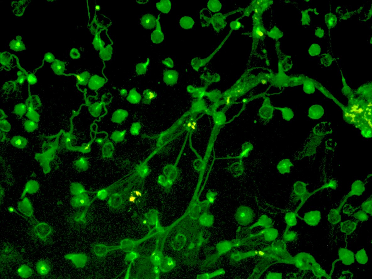
References:
- G. M. Arzumanyan and others., JRS, 2020, DOI: 10.1002/jrs.5844
- Arzumanyan G.M., et al., IJMS, 2023, DOI: 10.3390/ijms24065770