M. Arzumanyan, A. V. Bondarenko (BSUIR, Minsk, Belarus)
Z. Mamatkulov, E. Arynbek, D. S. Zakrytnaya, E. M. Demina, A. A. Shutikov, I. M. Pugachevskaya
The development of biosensing techniques for the reliable detection of various bioorganic molecules at ultralow concentrations is an urgent task for researchers. The discovery of SERS has paved the way for the detection of vital organic molecules from solutions in concentrations concerning single molecules, i.e. below 10-15 M. SERS is a powerful analytical instrument that allows to obtain specific information about the structure of molecules due to the registration of the enhanced Raman signal from plasmonic nanostructures - SERS-active substrates. The SERS effect is mainly provided by an anomalously strong electromagnetic field resulting from collective oscillations of conduction band electrons or plasmons excited on the surface of noble metal nanoparticles under laser radiation. It provides express analysis of various substances that allows to detect, identify and study the structure of target chemical compounds in ultra-low concentrations up to single molecules to solve problems in many areas of human life, such as medicine, biology, forensics, ecology, pharmaceuticals, and others. Of particular interest is the detection and investigation of bioorganic macromolecules using SERS, in particular, proteins with antiseptic properties, since they are promising for the development of nanomaterials for the prevention and treatment of bacterial and viral diseases.
The successful combination of the highly sensitive CARS Raman microspectrometer at FLNP with SERS-active substrates (Fig. 1) developed at BSUIR (Minsk, Belarus) have allowed to register the SERS spectra of bioorganic molecules adsorbed on silver-plated porous silicon (por-Si) from solutions with concentrations of 10-6 – 10-18 M (Fig.2).
Fig. 1. SEM images of SERS-active substrates based on (a, b) silver particles and (c, d) silver dendrites on porous silicon 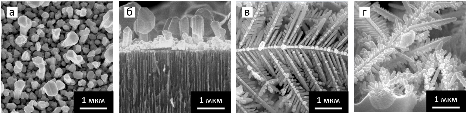
Fig. 2. SERS spectra of lactoferrin molecules adsoebed on silver particles and DTNB molecules on silver dendrites. 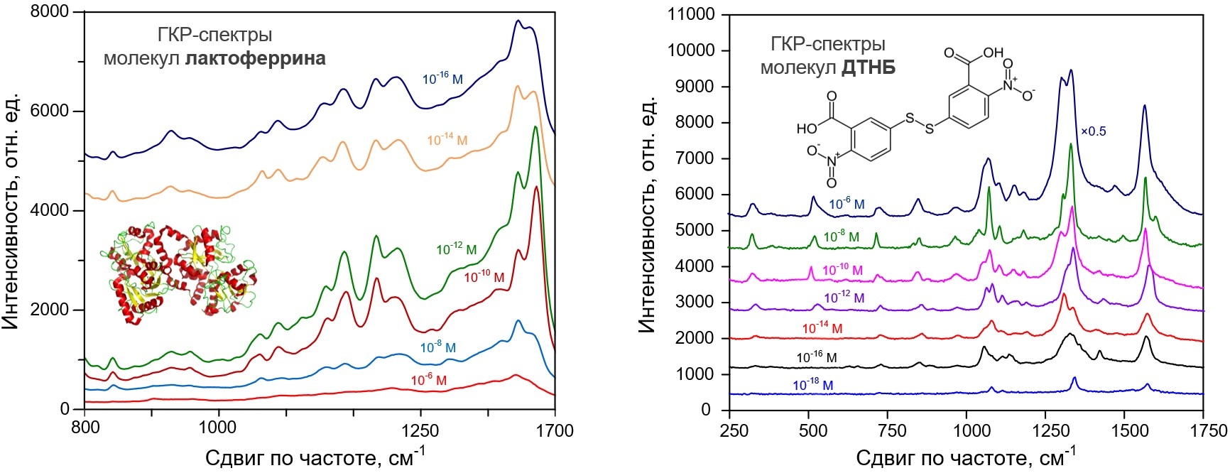
Publications:
- Zavatski, S., Khinevich, N., Girel, K., Redko, S., Kovalchuk, N., Komissarov, I., Lukashevich, V., Semak, I., Mamatkulov, K., Vorobyeva, M., Arzumanyan, G., Bandarenka, H. Surface Enhanced Raman Spectroscopy of Lactoferrin Adsorbed on Silvered Porous Silicon Covered with Graphene. Biosensors2019, 9, 34. https://doi.org/10.3390/bios9010034.
- Bandarenka, H. V., Khinevich, N. V., Burko, A. A., Redko, S. V., Zavatski, S. A., Shapel, U. A., Mamatkulov, K. Z., Vorobyeva, M. Yu. Arzumanyan, G. M. 3D Silver Dendrites for Single‐molecule Imaging by Surface‐enhanced Raman Spectroscopy. ChemNanoMat2020, 7, 2, 141- 149, https://doi.org/10.1002/cnma.202000521.